Ph.D. research & research software
TheraSeg
From multimodal segmentation research to an interactive imaging workflow.
A research platform bringing together report-guided SPECT/CT segmentation, multi-planar visualization, quantitative analysis, and structured export.
Graduate researcher & research software developer · CMIT Lab, WashU
January 2026 – Present
Research prototype
Associated segmentation method · retrospective study · 24 independent test patients.
01 / CONTEXT & CONTRIBUTION
TheraSeg: Automated Theranostics Segmentation & Quantification
Quantitative theranostic imaging needs organ and tumor regions that are consistent with the SPECT signal. The research challenge combines noisy emission images, anatomical mismatch, heterogeneous uptake, and limited tumor labels.
I am building TheraSeg to bring these methods into an interactive research workflow, with multimodal image review, segmentation overlays, adjustable parameters, and quantitative analysis.
My role in the work
- I am developing the platform and integrating organ/tumor segmentation with multi-planar visualization, quantitative analysis, and structured export.
- My methods work focuses on report-guided localization and unsupervised segmentation at CMIT Lab.
- I am the first author of the related segmentation conference abstract and presented this research and software at SNMMI events in 2026.
02 / METHOD & SYSTEM
From CT anatomy to SPECT contours.
01Establish anatomy from CT
TotalSegmentator provides initial CT organ masks. These anatomical priors define the starting regions for the SPECT workflow.
02Refine in emission space
CT-seeded region growing refines the organ regions against SPECT uptake, addressing the mismatch between anatomical boundaries and the emission image.
03Use clinical report context
An LLM identifies likely tumor-bearing anatomical categories from the clinical report, constraining the regions examined by the tumor segmentation stage.
04Inspect, quantify, and export
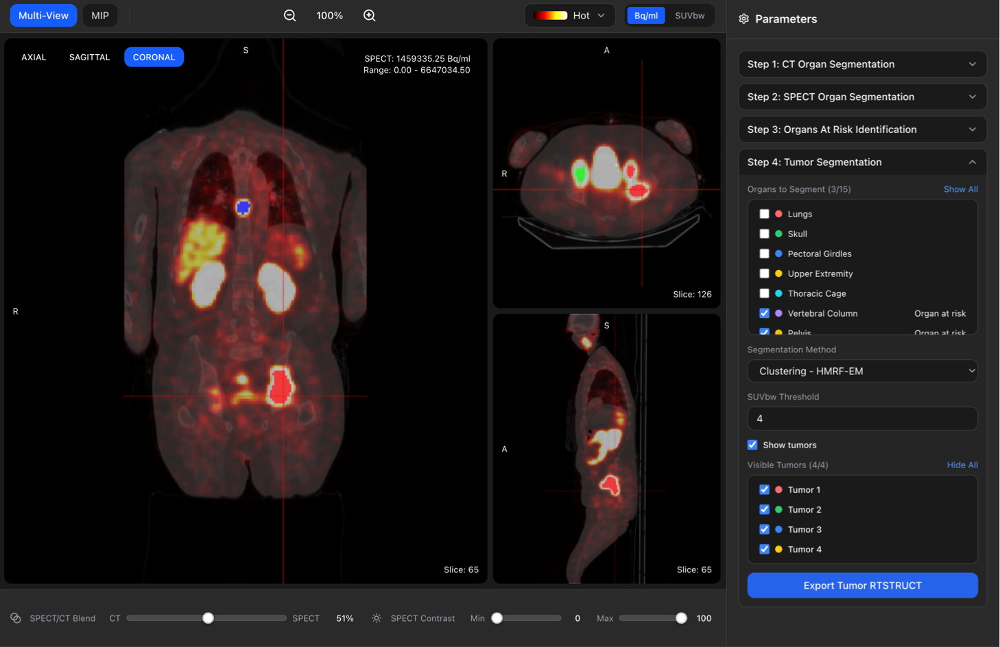
Linked anatomical views support review of HMRF-EM tumor segmentations, with controls for overlays, region selection, and parameters. Tumor contours can be exported as RTSTRUCT, alongside the platform’s quantitative analysis and structured-export workflow.
03 / RESULTS
Segmentation results and research software.
- TheraSeg brings fused SPECT/CT views and organ/tumor segmentation controls into a working research interface.
- We developed the method on 10 patients and tested it independently on 24. SPECT-space refinement increased mean organ Dice from 0.73 to 0.80, while mean tumor Dice was 0.43.
- The linked segmentation study includes the full method comparison, quantitative results, and per-patient examples.
What I take from this work
My goal with TheraSeg is to make segmentation methods easier to inspect, use, and connect to quantitative imaging research.
04 / DESIGN CHOICES
Connecting the method to an imaging workflow.
Refine anatomy in emission space
Directly transferring CT masks leaves a mismatch with SPECT uptake. In our independent test cohort, refining those masks in SPECT space increased mean organ Dice from 0.73 to 0.80. This comparison is central to the organ-segmentation workflow.
Constrain tumor localization with report context
The method uses clinical reports to select likely tumor-bearing anatomical categories, then applies unsupervised HMRF-EM segmentation within those regions. Mean tumor Dice of 0.43 shows that tumor delineation remains a harder problem than organ segmentation.
Let researchers inspect and export the contours
In TheraSeg, linked coronal, axial, and sagittal views place segmentation overlays beside their parameters. RTSTRUCT export connects the reviewed contours to subsequent imaging work; quantification and structured export are part of the platform I am developing.