CURRENT RESEARCH · Multimodal medical imaging
Report-guided segmentation for theranostic SPECT/CT
Can anatomical images and clinical reports guide segmentation when labeled SPECT data are scarce?
Conference abstract · Journal of Nuclear Medicine · SNMMI Annual Meeting · 2026 · First author · Current research
Results from our 2026 Oncologic Imaging Program (OIP) symposium abstract, covering 34 patients with 24 held out for independent testing. The related SNMMI conference abstract is linked above.
My contribution. I develop report-guided localization and unsupervised segmentation methods at CMIT Lab, and am first author of the related SNMMI abstract. I also develop TheraSeg to connect the methods to visualization and quantitative analysis.
01 / THE QUESTION
Background.
Post-therapy Lu-177 DOTATATE and PSMA SPECT/CT can support activity estimation and dosimetry, but quantitative analysis depends on identifying organs and tumors. Noisy, low-resolution SPECT images, heterogeneous tracer uptake, and variable lesion locations make this difficult.
CT supplies anatomical structure, yet acquisition-time misalignment can make direct CT-mask transfer inaccurate in SPECT space. Limited task-specific reference masks also constrain supervised segmentation training. This study combines anatomical priors, emission information, and clinical report context in an unsupervised tumor segmentation framework.
02 / THE APPROACH
Method, step by step.
Open a step to explore the approach.
01Establish anatomical priors
Pretrained TotalSegmentator generates initial organ masks from low-dose CT, providing anatomical guidance for the SPECT segmentation workflow.
02Refine in SPECT space
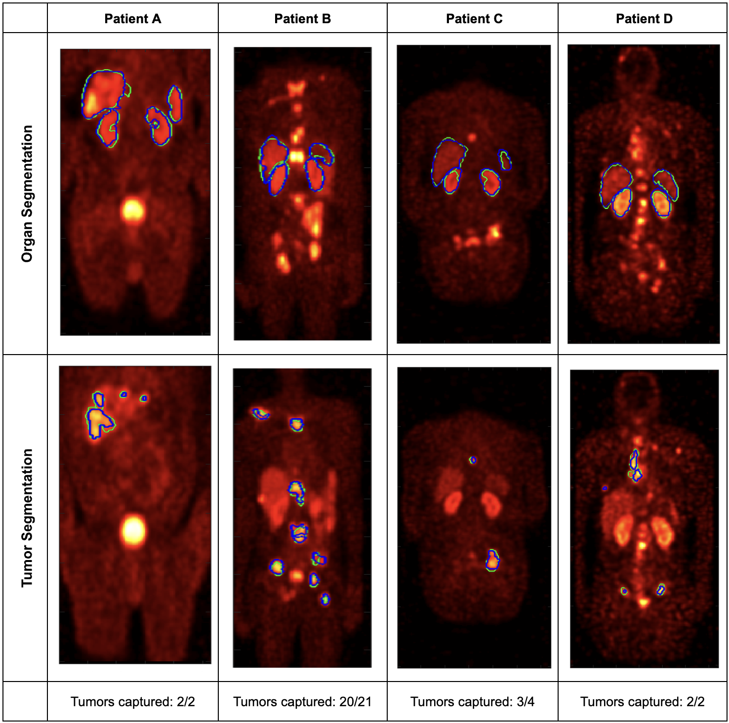
A CT-seeded region-growing method refines organ masks using SPECT uptake, addressing mismatch between CT anatomy and the emission acquisition. Refined masks are merged into clinically relevant anatomical categories.
03Use report context
A HIPAA-compliant language model parses the clinical radiology report to identify likely tumor-bearing anatomical categories. The report guides where tumor segmentation is performed.
04Segment and evaluate
Within the selected regions, hidden Markov random field–expectation maximization (HMRF-EM) and threshold-guided component selection identify tumors. Physician-defined contours provide the reference for evaluation.
Study design
Retrospective cohort: 34 patients; development: 10; independent test: 24. Organ refinement is compared with direct CT-mask transfer. Tumor baselines include 42% SUVmax thresholding, Otsu thresholding, gradient edge-based segmentation, and active contours.
03 / THE EVIDENCE
Results.
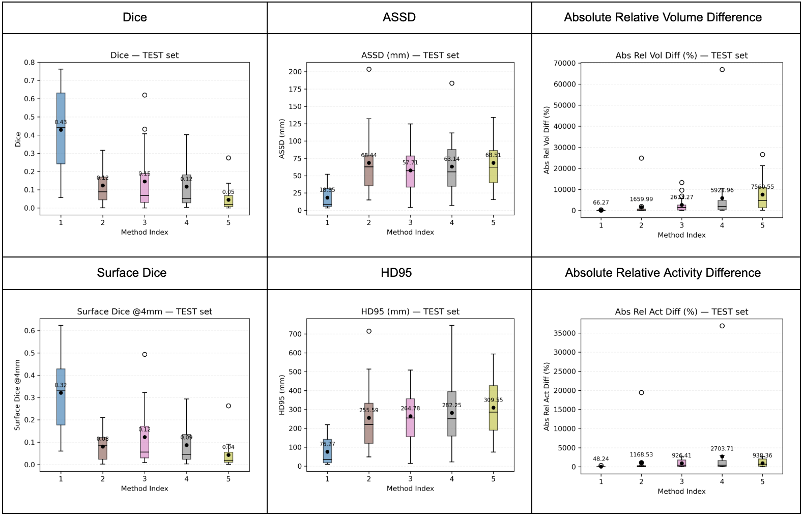
SPECT-space refinement improved organ overlap, boundary accuracy, and quantitative agreement. Our tumor segmentation method outperformed the conventional baselines we tested, although tumor segmentation remained substantially more challenging than organ segmentation.
CT-only masks versus SPECT-refined masks
Independent test cohort. Higher overlap is better; lower boundary distances and absolute relative differences are better. Surface Dice tolerance: 4 mm.
Mean organ Dice · scale 0–1
0 1
| Metric | CT-only | SPECT-refined |
|---|---|---|
| Mean Dice ↑ | 0.73 | 0.80 |
| Surface Dice at 4 mm ↑ | 0.41 | 0.48 |
| ASSD ↓ | 4.86 mm | 3.93 mm |
| HD95 ↓ | 13.58 mm | 10.96 mm |
| Absolute relative volume difference ↓ | 27.98% | 17.56% |
| Absolute relative activity difference ↓ | 20.04% | 13.08% |
Tumor segmentation on the independent test cohort
Tumor metrics on the independent test cohort, shown as mean ± standard deviation.
| Metric | Proposed method |
|---|---|
| Dice ↑ | 0.43 ± 0.21 |
| Surface Dice at 4 mm ↑ | 0.32 ± 0.15 |
| ASSD ↓ | 18.35 ± 16.44 mm |
| HD95 ↓ | 76.27 ± 71.12 mm |
| Absolute relative volume difference ↓ | 66.27 ± 73.75% |
| Absolute relative activity difference ↓ | 48.24 ± 75.10% |
Numerical results: 2026 OIP symposium abstract, Results section.
04 / WHAT IT MEANS
Conclusions.
- Anatomical priors become more useful for quantitative SPECT analysis when they are refined in emission space.
- Clinical report context offers a way to constrain tumor localization without requiring a task-specific supervised tumor segmentation model.
- The framework may support more reproducible activity estimation and downstream theranostic dosimetry workflows.
Limitations & next steps
We evaluated the method retrospectively on 34 patients, with 24 held out for independent testing. Moderate tumor Dice and variable quantitative errors motivate further validation on larger cohorts and in prospective studies.
05 / READ FURTHER
Citation & sources.
A Large-Language Model-Guided Unsupervised Segmentation Method for Lu-177 DOTATATE/PSMA SPECT/CT Images
Journal of Nuclear Medicine · SNMMI Annual Meeting · 2026;67(Supplement 1):261107
Citation
Chaurasia U, Abu-Mahfouz A, Rangbulla M, Qi K, Laforest R, Prasad V, Jha AK. A Large-Language Model-Guided Unsupervised Segmentation Method for Lu-177 DOTATATE/PSMA SPECT/CT Images. Journal of Nuclear Medicine. 2026;67(Supplement 1):261107. https://jnm.snmjournals.org/content/67/supplement_1/261107
Results from our 2026 Oncologic Imaging Program (OIP) symposium abstract, covering 34 patients with 24 held out for independent testing. The related SNMMI conference abstract is linked above.